How to Calibrate an Ocular Micrometer
Learn how to calibrate an ocular micrometer for precise microscopy measurements. This practical guide covers required tools, calculation methods, verification across magnifications, and documentation to ensure repeatable accuracy.

You will calibrate an ocular micrometer by aligning it with a stage micrometer, computing the exact micron-per-division value, and verifying accuracy at your working magnifications. Gather a stage micrometer, an eyepiece micrometer, a precision ruler or calipers, and a stable microscope setup before you begin. Document each step and record your results for repeatability.
Why calibrate an ocular micrometer
Calibrate Point's field guides emphasize that precise microscopic measurements start with a reliable reference. If you’re wondering how to calibrate an ocular micrometer, the answer is to establish a known scale in your eyepiece and translate divisions into real units. This ensures that measurements you make—whether counting cells, particles, or features—are consistent across sessions and magnifications. Regular calibration minimizes drift caused by lens wear, illumination changes, or aging equipment, and helps you compare results between labs or projects.
In practice, calibration aligns your eyepiece graticule with a stage micrometer so that each ocular division corresponds to a known distance in micrometers. By repeating this process when you change objectives, you preserve measurement integrity. The goal is not to memorize a single value, but to maintain a reliable conversion factor that applies under your usual lighting and temperature conditions. A correctly calibrated ocular micrometer becomes the backbone of quantitative microscopy, enabling accurate size estimates for routine work or formal analyses.
Core concepts: stage vs ocular micrometers
Two reference scales anchor calibration: the stage micrometer, which has a precisely etched ruler with known intervals, and the ocular micrometer, the grid etched inside the eyepiece. The core idea when learning how to calibrate an ocular micrometer is to determine how many micrometers correspond to a single ocular division at a given magnification. The conversion factor depends on magnification and objective, so you will develop a per-magnification chart. Because the ocular scale is part of the eyepiece and can vary between lenses, performing calibration for each objective you use yields the most accurate results. In practice, you align the scales as you would align a ruler with a caliper: bring the stage micrometer into view, count the ocular divisions across a known stage distance, and compute the conversion ratio. Once established, this ratio lets you estimate real sizes directly from ocular readings, a cornerstone of quantitative microscopy and image analysis.
Tools and setup for reliable calibration
A robust calibration requires clean, stable optics and a controlled environment. Before you begin, ensure the microscope is on a vibration-free bench, illumination is stable, and the eyepiece is free of dust. The essential tools include a stage micrometer slide with known distances, the ocular micrometer installed in the eyepiece, a precision ruler or calipers accurate to at least 0.01 mm, and a means to record measurements (lab notebook or digital log). If you use a camera or image analysis software, it can help capture and measure the lines more precisely, but manual counting remains perfectly valid. Label your calibration session with the objective lens you’re using, as the conversion factor will vary with magnification. For repeatability, keep a dedicated calibration slide and a dedicated eyepiece; mixing slides or epieces between sessions can introduce small errors. Finally, verify the visibility of the stage lines under your chosen illumination and ensure the ocular lines align crisply with minimal parallax for accurate counting.
A precise, repeatable calculation method
The fundamental calculation to answer how to calibrate an ocular micrometer is straightforward: micron per ocular division = (known stage distance in micrometers) / (number of ocular divisions that cover that distance). In practice you translate a measured stage distance into micrometers and divide by how many ocular divisions span that distance. Do this at your working magnification, then repeat with a second known distance to confirm consistency. If you observe a discrepancy, re-check alignment, focus, and parallax, then adjust the offset or stage zero as needed. Record both results and compare; the average of multiple determinations is your calibrated factor for that magnification. You can extend this method to multiple objective lenses by repeating the same procedure for each objective; plotting the resulting micron-per-division values against magnification creates a simple calibration curve you can reference during measurements. Document any instrument changes, such as eyepiece replacement or adjustments to the camera, since these can alter the scale slightly.
Verification across magnifications
After computing the conversion factor for a given objective, test it by measuring a second known distance on the stage micrometer and check whether the ocular reading translates to the expected micron distance. Repeat measurements at 4x, 10x, 40x, and 100x objectives if available, noting any deviations. A robust calibration will show minimal variation between trials, ideally within a small, predefined tolerance you set during planning. If variations exceed tolerance, revisit alignment, parallax, and illumination until readings stabilize. It's common to run a quick verification sequence at the end of each calibration session to confirm that the most recent value remains valid. Keep a log of these checks so you can detect gradual drift over time and schedule a re-calibration before it significantly affects your work.
Documentation, storage, and rechecking
Record the key numbers: magnification, stage micrometer division size, ocular micrometer divisions observed, and the calculated micron-per-division value. Include the date, operator, instrument model, and any environmental conditions that might influence results. Store a copy of the calibration slide for reference. Use a dedicated notebook page or digital record that you can update when changes occur (new eyepieces, lamps, or cameras). Schedule periodic rechecks—some labs calibrate monthly, others quarterly depending on usage. If you notice systematic drift, re-calibrate with the same procedure and compare with prior results to quantify the drift direction and speed. Finally, share the calibration data with teammates or collaborators to ensure that everyone uses the same scale in measurements, which is essential for reproducibility and data integrity.
Tools & Materials
- Stage micrometer slide(0.01 mm divisions; verify accuracy before use)
- Ocular micrometer / eyepiece micrometer(Ensure proper installation and alignment with the eyepiece)
- Precision ruler or calipers(0.01 mm resolution recommended for cross-checks)
- Calibration log notebook or digital log(Record magnification, measurements, and results)
Steps
Estimated time: 30-60 minutes
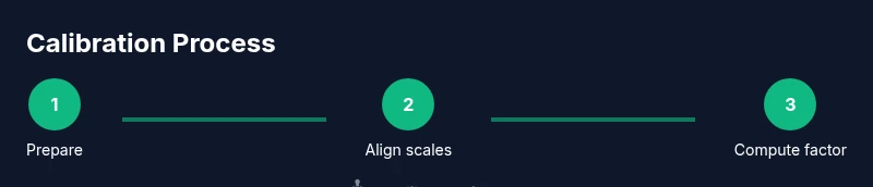
- 1
Prepare and inspect the setup
Power on the microscope, clean the objective and eyepiece surfaces, and ensure stable illumination. Inspect the stage micrometer for clarity and the ocular micrometer for proper zero alignment. This step reduces parallax errors and ensures reliable counting.
Tip: Remove dust with a soft brush and avoid touching optical surfaces. - 2
Install references and verify zero
Insert the stage micrometer slide and align the eyepiece micrometer so the zero lines coincide. Lock the stage if your instrument has a lock to prevent drift during measurements.
Tip: Double-check that the stage zero lines up exactly with the ocular zero before counting. - 3
Focus and count ocular divisions
Bring the stage micrometer into view at the chosen magnification and count how many ocular divisions span a known stage distance. Maintain a steady viewpoint to avoid parallax error during counting.
Tip: Look straight on to minimize parallax; count slowly and pause between counts to reduce mistakes. - 4
Compute micron-per-division value
Apply the formula: micron per ocular division = (stage distance in micrometers) ÷ (ocular divisions across that distance). Repeat with a second distance to confirm, and record both results.
Tip: Use exact fractions or calculator to reduce arithmetic errors. - 5
Verify across a second magnification
Repeat the same measurements for a different objective lens or magnification to check consistency. Ideally the conversion factor remains stable within your predefined tolerance.
Tip: Document any observed drift between magnifications. - 6
Document and archive results
Log the magnification, dates, instrument model, and the calculated micron-per-division value. Store a copy of the slide and notes for future reference and rechecks.
Tip: Create a simple calibration record template for quick future updates.
Questions & Answers
What is an ocular micrometer?
An ocular micrometer is a calibrated scale embedded in the microscope eyepiece. It requires calibration against a stage micrometer to provide real size values in micrometers.
An ocular micrometer is a built-in measuring scale in the eyepiece that needs calibration against a stage micrometer.
Why calibrate at multiple magnifications?
Calibration values can vary with magnification and objective. Calibrating for each setup ensures consistent size estimates across studies and sample types.
Calibration values can change with magnification, so calibrate for each setup.
Can I reuse a calibration across different objectives?
Calibration values are typically specific to a magnification-objective combination. Re-check whenever you switch objectives or eyepieces.
Usually you need new calibration for different objectives or eyepieces.
What should I do if measurements don’t match?
Recheck alignment, ensure there is no parallax, and re-run the calibration with fresh references. If issues persist, escalate to re-calibration.
If measurements don’t match, redo alignment and re-check the scales.
How often should recalibration occur?
Set a practical schedule based on workload and instrument changes; monthly or quarterly are common in busy labs.
Calibrate on a schedule that fits your workflow, especially after changes.
Is software necessary for calibration?
Software can help measure lines on captured images, but manual counting with a stage micrometer is widely used and reliable. Software is optional.
Software can help, but you can calibrate using manual counts.
Watch Video
Key Takeaways
- Calibrate for each magnification used.
- Align stage and ocular scales precisely.
- Document all parameters and results.
- Verify with multiple known distances to confirm accuracy.